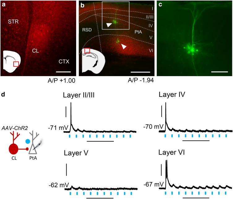
Fig. 3.
Claustrum afferent stimulation drives PtA neuron firing in layers II/III, IV, and VI but not V. a Photomicrograph showing AAV-ChR2 (red) expression after injection into the claustrum. The photomicrograph location is indicated by the red box on the cartoon. b Photomicrograph showing PtA layer-specific innervation of claustrum afferents expressing ChR2 (red box shows PtA inset region). Neurons across PtA layers were targeted for whole-cell recordings and filled with neurobiotin (green, arrowheads). Cortical layers were delineated by staining with 4′,6-diamidine-2′-phenylindole (DAPI, not shown). Innervation of layer V was relatively sparse compared to other cortical layers. c Inset from b showing PtA layer V pyramidal neuron at high magnification. d Left: schematic showing claustrum innervation of PtA layers. Right: representative traces showing responses to optogenetic stimulation of claustrum afferents (20 Hz, 470 nm light). Action potential firing was noted at initial light pulses across PtA layers except for layer V. Horizontal scale bars—200 µm a; 400 µm b; 100 µm c; 200 ms d. Vertical scale bars—30 mV. A/P anterior/posterior, RSD retrosplenial dysgranular cortex

