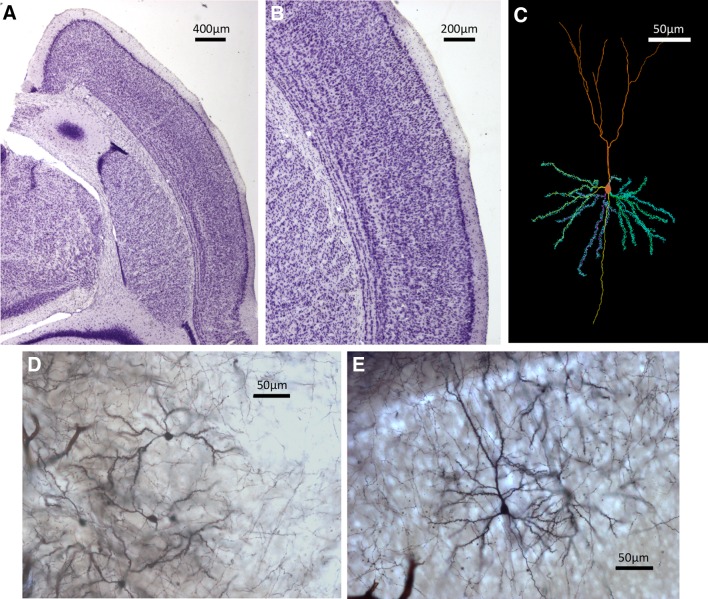
Fig. 1.
Examples of histological sections and neuron tracings. a Nissl-stained section of a shrew brain hemisphere showing a dorsal medial area. b Details of a Nissl-stained section depicting a portion of the neocortex. c Neuron tracing as depicted by Neurolucida. d Medium spiny neurons in the caudoputamen stained with the Golgi technique. e Pyramidal neurons in the neocortex

