Fig. 3.
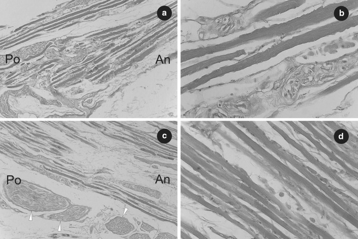
Histological observations on the right accessory rectus muscle. a Part of the superior head of the right accessory rectus muscle. Fibers of striated skeletal muscle have been visualized. H&E stain, ×10 objective. b Sample fibers of striated skeletal muscle obtained from the superior head of the right accessory rectus muscle. The striations of skeletal muscle tissue have been visualized. H&E stain, ×40 objective. c Part of inferior head of the right accessory rectus muscle shown near its origin from the inferior rectus muscle. Fibers of striated skeletal muscle as well as cross sections of small nerves (marked by white arrowheads) have been visualized. H&E stain, ×10 objective. d Sample fibers of striated skeletal muscle obtained from the inferior head of the right accessory rectus muscle. The striations of skeletal muscle tissue have been visualized. H&E stain, ×40 objective. An anterior, Po posterior

