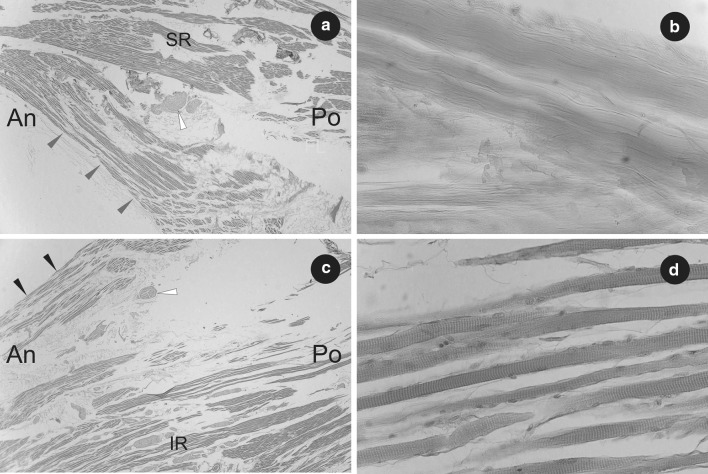
Fig. 4.
Histological observations on the left accessory rectus muscle. a Part of the superior head of the left accessory rectus muscle (marked by grey arrowheads) shown at its origin from superior rectus muscle (SR). H&E stain, ×2 objective. Fibers of striated skeletal muscle and cross-section of small nerve (marked by white arrowhead) have been visualized. b Part of the tendon with visible bundles of collagen fibers (tissue sample taken from the origin of the left accessory rectus muscle). H&E stain, ×40 objective. c Part of inferior head of the left accessory rectus muscle (marked by black arrowheads) shown at its origin from the inferior rectus muscle (IR). H&E stain, ×2 objective. Fibers of striated skeletal muscle and cross section of small nerve (marked by white arrowhead) have been visualized. d Sample fibers of striated skeletal muscle obtained from the inferior head of the left accessory rectus muscle. The striations of skeletal muscle tissue have been visualized. H&E stain, ×40 objective. An anterior, Po posterior

