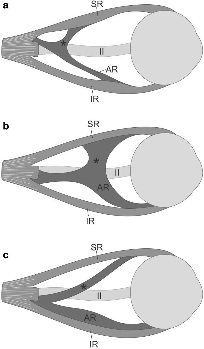
Fig. 5.
Different variants of the supernumerary orbital muscles reported in the literature. Lateral view. For ease of comparison and increased transparency, the same side has been presented on all schemes. Terminology applied by von Lüdinghausen et al. [17] has been taken into account. a Anatomical variation described in this report—an accessory muscle observed on 68-year-old cadaver with no eye movement abnormalities reported in the medical history. The accessory muscle is divided into two delicate slips (heads): superior (marked by black asterisk)—forming muscular bridge connected to the superior rectus muscle (SR); and inferior—corresponding to accessory rectus muscle (AR) and attached in the anterior half of the inferior rectus muscle (IR). b Supernumerary orbital muscle (AR) with a broad muscular bridge (marked by black asterisk) to the SR and attachment to the anterior part of the IR. The accessory muscle was well-separated from the IR. c Supernumerary orbital muscle (AR) with a thin muscular bridge (marked by black asterisk) to the SR and close attachment to the anterior part of the IR. Variants b and c were described by von Lüdinghausen et al. [17] on the adult cadaver with no problems with mobility of the eyeball in the medical history. II optic nerve

