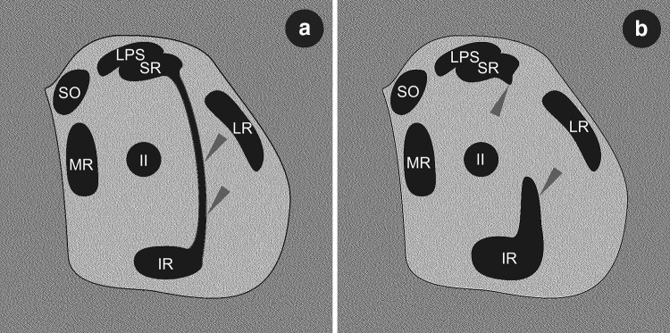
Fig. 6.
Schematic drawings simulating MRI or CT coronal scans demonstrating spatial organization of muscular structures within the orbit in the event of presence of accessory (supernumerary) rectus muscles or muscular bands between superior and inferior rectus muscles. The drawings have been prepared on the basis of comparison of different MRI scans presented by Khitri and Dremer [7] and Kightlinger at al. [8]. a Complete muscular bridge seen between temporal edges of superior and inferior rectus muscles (marked by grey arrowheads). On drawing (b) only fragments of certain heads of the supernumerary rectus were captured (grey arrowheads). IR inferior rectus muscle, LR lateral rectus muscle, LPS levator palpebrae superioris muscle, MR medial rectus muscle, SR superior rectus, SO superior oblique muscle, II optic nerve

