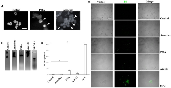
Figure 3.
Trophozoites induces NETosis without evidence of apoptosis or necrosis. (A) Neutrophils were incubated with 20 nM PMA or trophozoites (ratio 20:1). After 4 h of incubation, cells were fixed and stained with DAPI. Decondensed chromatin is shown (arrowheads). Images were taken at 100x magnification. Scale bar 10 μm. (B) DNA from neutrophils (1 × 10⁶) treated for 1 h with trophozoites (5 × 10⁴) or 20 nM PMA or 10 μM A23187 or heat (50°C) were extracted and run in 1.8% agarose gel and the bands visualized by staining with ethidium bromide. (C) Phosphatidylserine (PS) exposition was assessed by fluorescence microscopy using FITC-annexin V. (D) Percentage of cells positive to PS was determined in a total of 300 stained neutrophils. Values are means ± SD of three independent experiments. *p < 0.001.

