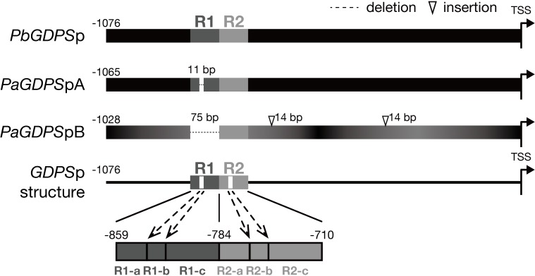
FIGURE 1.
Promoter structure of GDPS. Promoter structure of GDPS was revealed by the comparison of three sequences of PbGDPSp, PaGDPSpA, and PaGDPSpB. Two repeats located from –859 to –710 of PbGDPSp were named as R1 and R2. The repeat was further dissected into three subunits based on 11-bp deletion located in the center of R1. This deletion was referred as R1-b, and the sequences prior to and behind R1-b was R1-a and R1-c, respectively. The corresponding dissection in R2 was R2-a, R2-b, and R2-c. TSS indicates the translation start site (ATG). Black color gradient in PaGDPSpB indicated its numerous substitutions compared to PbGDPSp.

