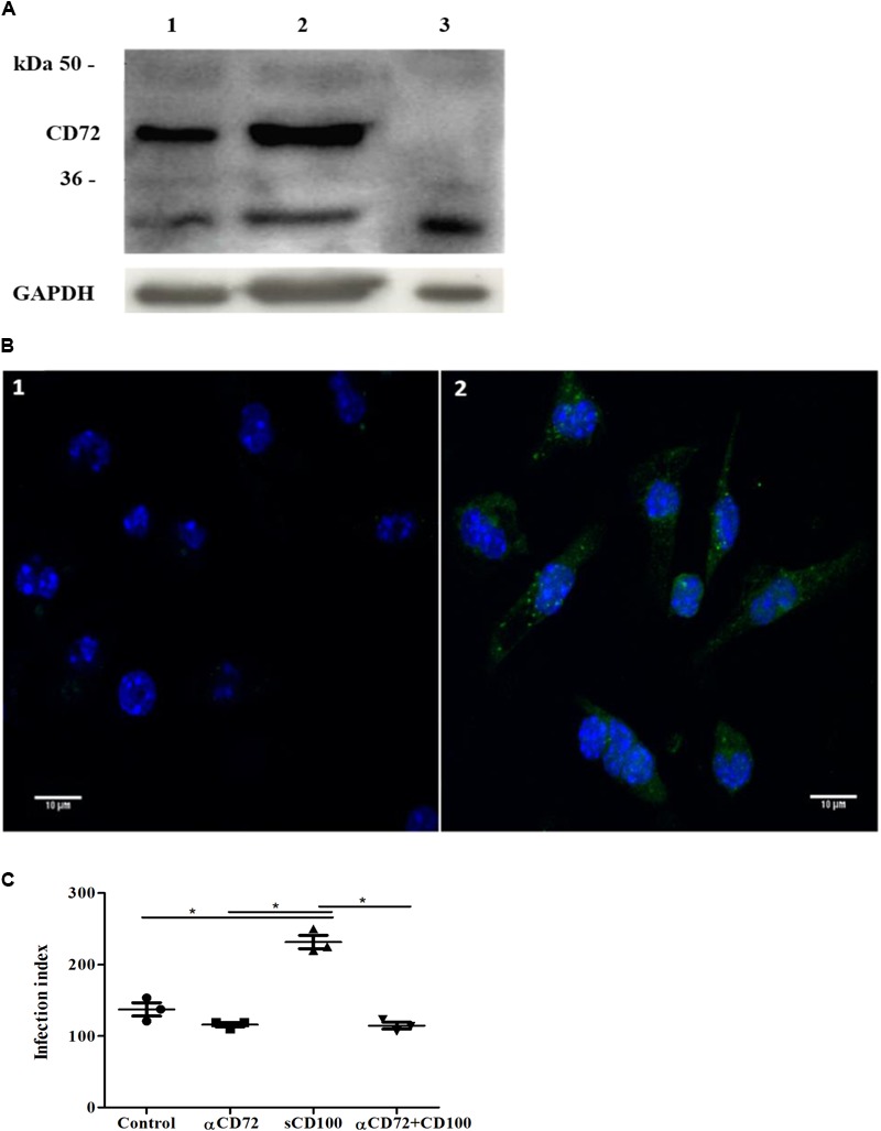
FIGURE 5.

Expression of CD72 and its role in sCD100 effects on macrophages. (A) Western blot showing expression of CD72 and GAPDH in peritoneal macrophages from BALB/c mice (lane 1), macrophage incubated with 200 ng/mL of sCD100 for 48 h (lane 2) and L929 cells (negative control - lane 3). Thirty micrograms of protein extracts were analyzed in 10% SDS–PAGE. (B) Immunofluorescence staining for CD72 in peritoneal macrophages from BALB/c mice: control incubated with anti-rabbit Alexa Fluor 488 secondary antibody and DAPI (image 1), macrophages incubated with anti-CD72, anti-rabbit Alexa Fluor 488 secondary antibody and DAPI (image 2). Images were captured in Zeiss LSM 780-NLO confocal microscope, magnifying 63 ×, 1.0 zoom. (C) Infection index of macrophages from BALB/c mice in the presence or absence of sCD100, preincubated or not with anti-CD72 for 48 h. Data represent means and standard deviations of one experiment with triplicates. Statistical analysis was performed by ANOVA followed by Tukey’s post-test, and significant differences are labeled as ∗p ≤ 0.05.
