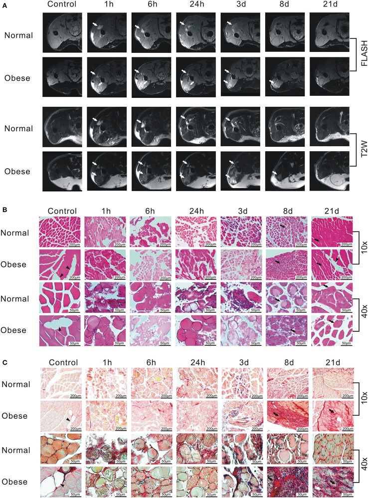
Figure 3.
Monitoring of the regeneration process after induction of a blunt muscle injury in female normal weight and obese C57BL/6J mice. (A) FLASH and T2W-RARE recordings of two female C57BL/6J mice receiving either ND or HFD. Arrow = edema. Same animals were used for the whole time course. Prescan (Control) did not show any signs of injury, whereas an edema can be seen starting 1 h post-injury. Edema formation increases until 6 h post-injury, then recedes within 21 days, whereas the obese mouse shows signs of edema 8 days after injury. The normal weight mouse seems to recover within the observed time frame. (B) Hematoxylin-eosin (HE) staining of muscle tissue sections. The staining shows damaged muscle, inflammatory cells and subsequent myofiber regeneration within 21 days post-injury. Triangle = fat inclusion, arrow = newly regenerated myofiber. (C) Sirius Red staining of muscle tissue sections. Triangle = fat inclusion, arrow = fibrosis. Pictures were taken with Olympus IX81 using Xcellence v.1.2. Scale = 200 μm at 10x magnification; Scale = 50 μm at 40x magnification.

