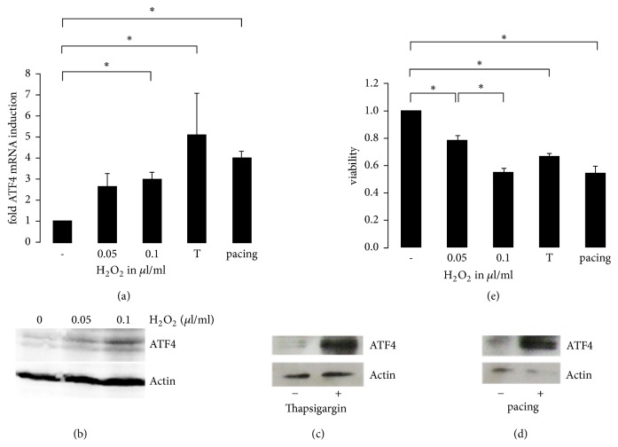
Figure 1.
ATF4 expression in H 2 O 2 and thapsigargin treated cardiomyocytes and in electrically stimulated cardiomyocytes. (a) Real-time PCR of ATF4 mRNA expression in cultured HL-1 cardiomyocytes in response to treatment with 0, 0.05, or 0.1 μl/ml H2O2 (n = 3), real-time PCR of ATF4 mRNA expression in cultured cardiomyocytes in response to treatment with 1 μM thapsigargin (n = 3), and real-time PCR of cardiomyocytes stimulated electrically with 4 Hz for 20 h (n = 3). Representative western blots showing protein expression of ATF4 in response to H2O2 treatment (b) and in response to thapsigargin treatment (c). (d) Cardiomyocytes were electrically stimulated with 4 Hz for 20 h and ATF4 protein expression was detected. (e) MTT assay displaying viability of cardiomyocytes treated with H2O2 (n = 3), 1 μM thapsigargin (n = 3), and paced HL-1 cardiomyocytes (n = 5). ∗ indicates p < 0.05.

