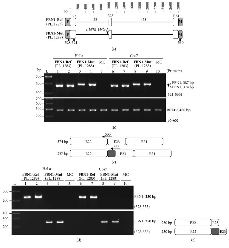
Figure 2.
RT-PCR analysis of minigene-derived transcripts. (a) Schematic representation of FLAG (F) and Myc- (M-) tagged reference (FBN1-Ref) and mutant (FBN1-Mut) minigene plasmids (PL). Exons (E) are denoted with white boxes and introns (i) with solid black horizontal lines. The approximate location of the primers for downstream RT-PCR analysis is shown (for primer sequences see Table 1). (b) Expression of FBN1-Ref and FBN1-Mut minigenes in HeLa and COS-7 cells as revealed by RT-PCR, using primers (521 and 530) targeting E22 and Myc. A representative of two independent experiments for each transfection is shown. RPL19 amplification was carried out as an input RNA control for the RT-PCR. L: size reference ladder. MC: mock cells. (c) Schematic representation of PCR products corresponding either to correct splicing (374 bp band) or to partial inclusion (dark grey box) of intron 22 (387 bp band) as revealed by sequencing (see Supp. Figure S3). (d) Partial inclusion of intron 22 in transcripts was assayed by using different combinations of primers targeting FLAG (primer 528 shown in (a)), exon 22/exon 23 junction (primer 533), or exon 23/retained intron 22 inside (primer 535). (e) A diagram of the PCR products (i.e., the 230 bp and 250 bp bands shown in (d)) as revealed by sequence analysis.

