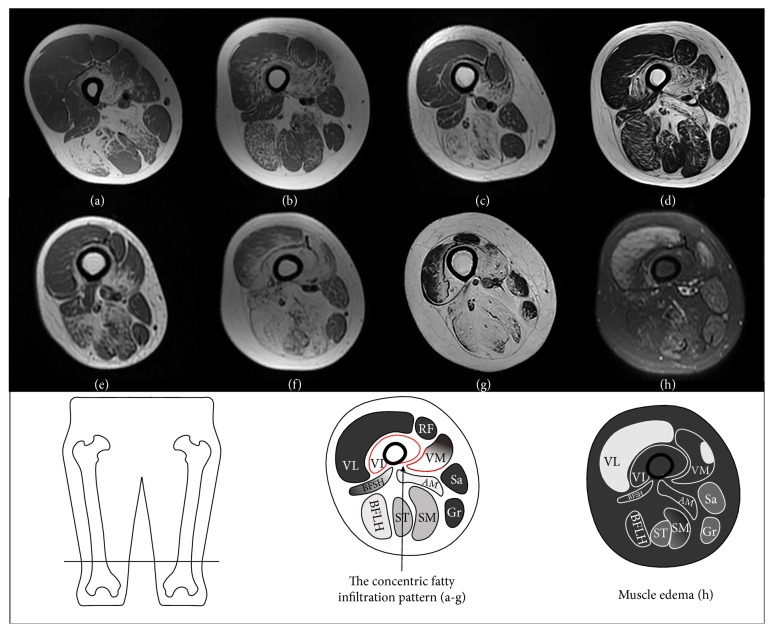
Figure 2.
Axial T1-weighted images of the right thigh for patients 4–10 (a–g) showing a concentric fatty infiltration pattern around the distal femoral diaphysis and an axial short T1 inversion recovery image of the right thigh in patient 9 (h) showing relatively marked edematous changes in the thigh muscles.

