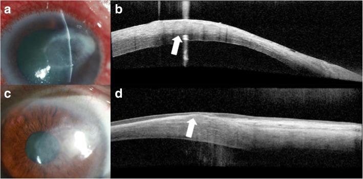
Fig. 13.
Slit lamp photograph and AS-OCT of infectious keratitis and subsequent corneal scarring. a Slit lamp photograph of a patient with contact lens related Pseudomonas infectious keratitis. b AS-OCT shows diffuse stromal hyperreflectivity and thickening in the area of the infiltrate involving nearly 50% of the stroma (arrow). c Slit lamp photograph of a compact, subepithelial scar after infectious keratitis. d AS-OCT shows subepithelial thinning and hyperreflectivity in the area of the corneal scar (arrow)

