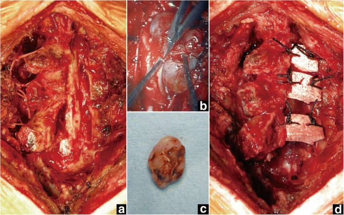
Fig. 2.
Case 1. Intraoperative photographs. a Laminae from C5 to C7 are fully opened with the right side as the opening side. b After opening the dura, the tumor is detached from the spinal cord microscopically. c The tumor is totally extirpated. d After extirpation of the tumor and dural closure, HA spacers are placed between the right-side laminae and lateral mass from C5 to C7

