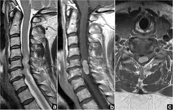
Fig. 4.
Case 2. Preoperative magnetic resonance imaging (MRI) of the cervical spine. a Sagittal T2-weighted MRI shows the intradural extramedullary spinal cord tumor with heterogeneous signal hyperintensity at C5-C6 and spinal canal stenosis at C4-C7. b, c Gadolinium-enhanced sagittal MRI (b) and axial MRI (c) at the C5-C6 level show homogeneous enhancement of a tumor located dorsally and to the left of the spinal cord

