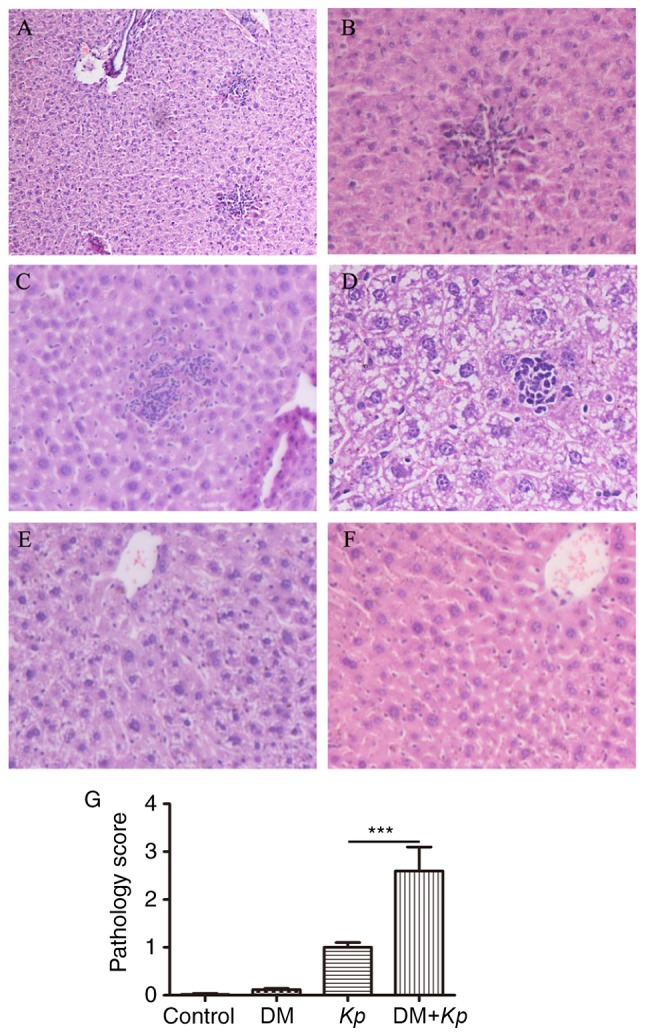
Figure 2.

Histochemical analysis of liver tissues. (A-C) Kp-infected mice with DM. The magnifications utilized for (A-C) are ×100, ×200 and ×200, respectively. (D) Kp-infected normal mice (magnification, ×400). (E) Kp-infected normal mice (magnification, ×200). (F) Control mice (magnification, ×200). (G) Semi-quantitative analysis of histological observations (magnification, ×200). ***P<0.001. Kp, Klebsiella pneumoniae; DM, diabetes mellitus.
