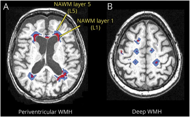
Figure 2. NAWM layer masks.

Red areas represent WMH. The light blue, blue, and white layers represent NAWM layer masks for (A) periventricular WMH and (B) deep WMH. The innermost layer adjoining WMH is NAWM layer 1 and the outermost layer away from the WMH is NAWM layer 5. NAWM = normal-appearing white matter; WMH = white matter hyperintensity.
