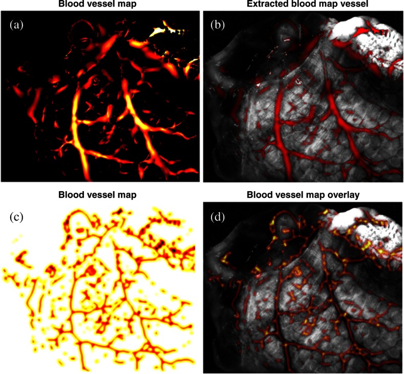
Fig. 4.
(a) Blood vessel segmentation using Frangi 2-D filter.24 (b) Blood vessel segmentation result (red) image overlay on the single-band image at 470 nm. (c) Blood vessel map created by Gaussian filter smoothing of the output of the Frangi 2-D filter. (d) Image overlay of inverted vessel map (inverted for better visualization) on the single-band reflectance image of the intestine.

