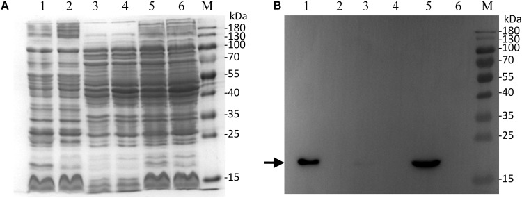
FIGURE 5.
Western blot detection and localization of RDD in Escherichia coli. Membrane fraction, cytoplasmic one and cell extract from E. coli KNabc/pET22b-PRO-RDD (Lanes 1, 3, 5) and KNabc/pET22b (Lanes 2, 4, 6) were sampled, respectively, and then subjected to SDS-PAGE (A) and western blot (B) analyses. The position of target protein RDD fused with an N-terminal His6 tag is shown with a solid arrow.

