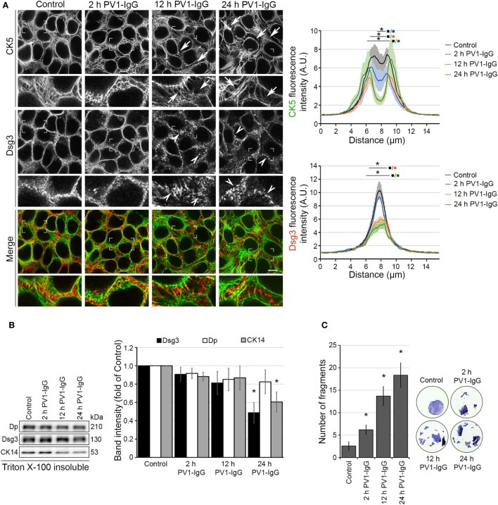
Figure 1.
Structural changes of human keratinocytes in response to pemphigus vulgaris (PV) antibody exposure. HaCaT keratinocytes expressing cytokeratin5 (CK5)-YPF (HaCaT-CK5) were incubated with PV1-IgG for 2, 12, or 24 h. Images shown are representatives of >3 independent experiments. (A) Desmoglein (Dsg)3 staining and CK5 expression in response to the antibody binding. Loss of keratin filaments in the cell periphery is marked by arrows and Dsg3 alterations by arrowheads. Comparison of fluorescence profiles on a 15 µm line perpendicularly to the membrane of two adjacent cells (n = 75 cells from three independent experiments, *p < 0.05 vs. control). Bar represents 10 µm. (B) Triton X-100 insoluble fraction, representing the cytoskeletal-bound fraction, of HaCaT-CK5 lysates after incubation with PV1-IgG for the indicated period of time (n = 6). Densitometric analysis of structure proteins shown as fold of control (n = 6, *p < 0.05 vs. control). (C) Dispase-based dissociation assays in HaCaT-CK5 keratinocytes after PV1-IgG treatment (n = 5, *p < 0.05 vs. control). Representative images of cell sheets after applied sheer stress stained with 10 µM MTT for better visibility.

