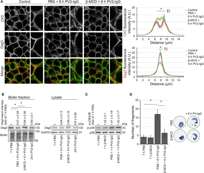
Figure 6.
Inhibition of Dsg3 internalization through cholesterol depletion did not prevent PV-IgG-driven keratin alterations. HaCaT-CK5 keratinocytes were pre-incubated with either PBS or β-MCD as a cholesterol-depleting agent for 1 h and then exposed to PV2-IgG for 6 h. Images shown (A) are representative for four independent experiments. For analysis a bar of 15 µm was applied perpendicularly over the cell border of two adjacent cells and the fluorescence intensity was plotted for both Dsg3 and CK5 (n = 100 cells from four independent experiments; *p < 0.05). Bar represents 10 µm. (B) Streptavidin pulldown of biotinylated membrane Dsg3 was performed with HaCaT-CK5 keratinocytes (n = 3–5, *p < 0.05). (C) Phosphorylation of p38MAPK was determined by densitometry. (D) Intercellular adhesion was determined by dispase-based dissociation assays in HaCaT keratinocytes (n = 8, *p < 0.05).

