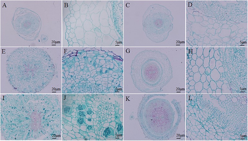
FIGURE 3.

Histocytological analysis of cross-sections of inoculated resistant and susceptible plants. (A,B,E,F,I,J) Sections of root of Brassica napus. (C,D,G,H,K,L) Sections of root of Matthiola incana. (A–D) Sections of inoculated roots at 14 days after inoculation (DAI). (E–H) Sections of inoculated roots at 20 DAI. (I–L) Sections of inoculated roots at 28 DAI. Adjustments for magnification and illumination were performed to allow optimal viewing of the individual sections.
