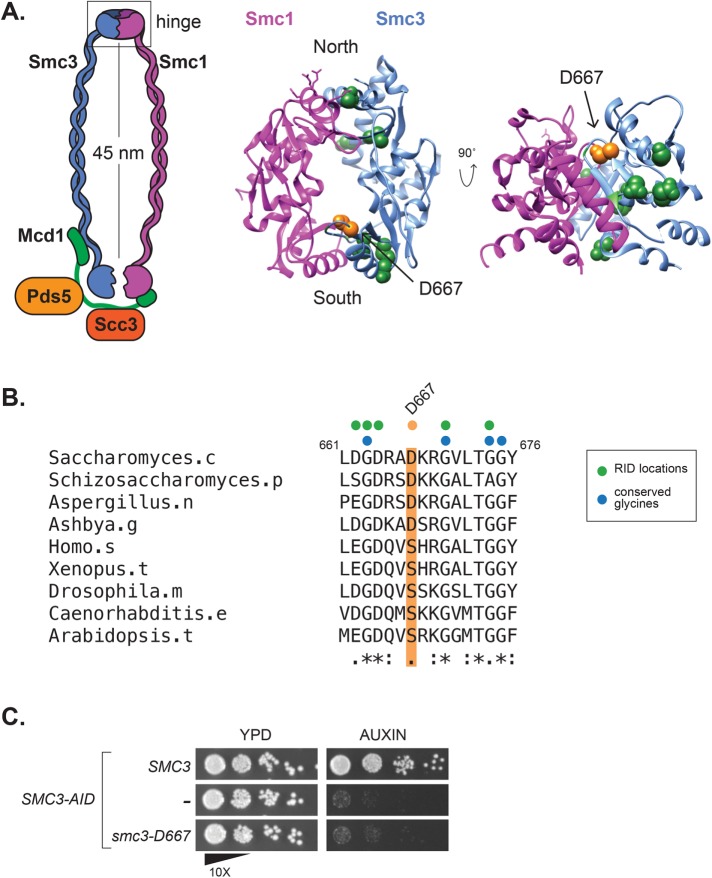
FIGURE 1:
The smc3-D667 RID mutation maps to a loop near the South interface of the Smc3p hinge. (A) Diagram of cohesin highlighting location of the smc3-D667 RID insertion. The homologous residue of smc3-D667, highlighted in orange, was determined by sequence alignment using ClustalW and mapped onto the mouse Smc1p/Smc3p hinge crystal structure (PDB: 2WD5; Kurze et al., 2011). Other RIDs isolated in this screen and located in the hinge domain are represented as green spheres, and their positions were also approximated by sequence alignment. (B) Sequence alignment of Smc3p homologues showing the conserved region around D667. The position of Asp667 is highlighted in orange, and the sequence of the five-amino-acid insertion, AAAAD, that follows Asp667 in the smc3-D667 RID is depicted above as an orange dot. The position of other RIDs in this region are shown with green dots and conserved glycine residues shown with blue dots. (C) The smc3-D667 allele under the native SMC3 promoter is unable to support viability. Cultures of haploid strains SMC3 SMC3-AID (BRY474), SMC3-AID (VG3651-3D), and smc3-D667 SMC3-AID (BRY482) were grown to saturation in YPD and then plated in 10-fold serial dilutions onto YPD alone (YPD) or containing 0.75 mM auxin (auxin) and then grown for 2 d at 23°C.

