FIGURE 2:
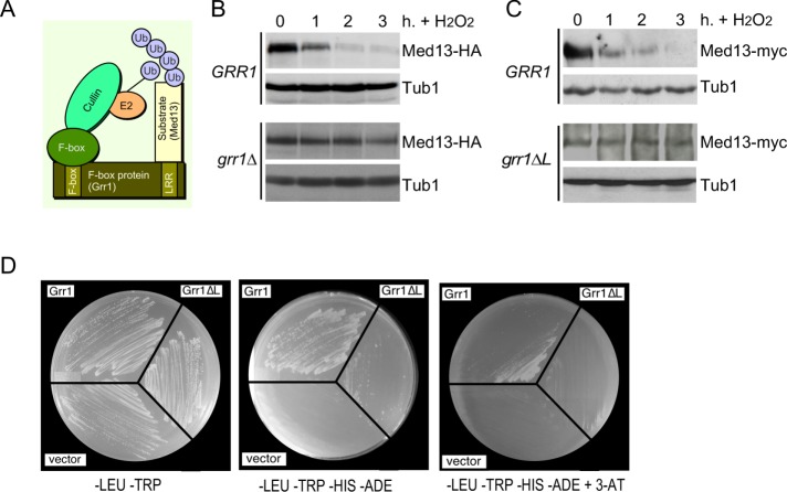
SCFGrr1 mediates Med13 degradation following H2O2 stress. (A) Model of the SCFGrr1. (B) Wild-type (RSY10) and grr1∆ cells (RSY1770) harboring Med13-HA (pKC801) were treated with 0.4 mM H2O2 for the time points indicated and Med13 levels were analyzed by Western blot. (C) Top panel: RSY1798 (MED13-myc::KAN) was treated with 0.4 mM H2O2 for the time points indicated and Med13 levels were analyzed by Western blot. Tub1 levels were used as loading controls. Bottom panel: RSY1771 (grr1∆::HIS3 MED13-myc::KAN) harboring ADH1PRO-grr1∆L was analyzed as for RSY1798. Tub1 levels were used as loading controls. (D) Yeast two-hybrid analysis of Med13 and Grr1 derivatives. Y69a cells harboring Med13-activating domain plasmid (pKC800) and either pAS2, pAS-Grr1, or pAS2-Grr1∆L binding domain plasmids were grown on –LEU, –TRP dropout medium to select for both plasmids (left panel) and on –TRP, –LEU, –HIS –ADE (middle panel), and –TRP, –LEU, –HIS, –ADE +3-AT (right panel) to test for Med13-Grr1 interaction.

