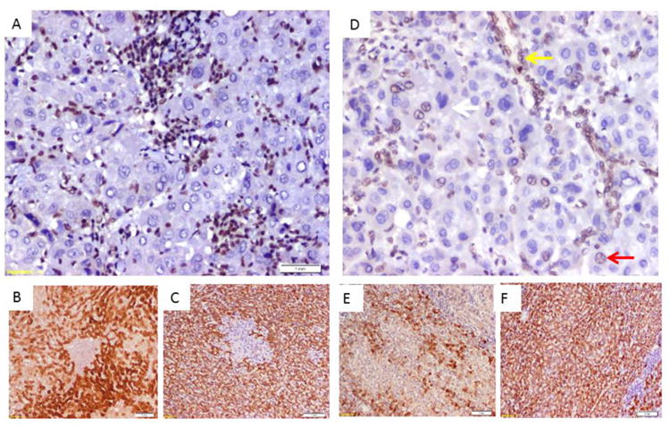
Figure 2. Immunostaining of HCC showing association of BAP1 loss with positive staining with bile duct cell marker cytokeratin 7.

A) HCC case with homogenous loss of nuclear staining of BAP1, note positive staining in stromal and infiltrating inflammatory cells. B) CK7 immunostaining of the tumor in (A) showing positive expression of tumor cells to the bile duct epithelial marker. C) HepPar immunostaining of the tumor in (A) showing positive expression of tumor cells to the hepatocyte marker.
D) HCC case with heterogenous loss of nuclear staining of BAP1(white arrow loss of nuclear expression, red arrow show preserved expression, yellow arrow show preserved expression in a non-tumor bile duct). E) CK7 immunostaining of the tumor in (D) showing heterogenous expression of tumor cells to the bile duct epithelial marker. C) HepPar immunostaining of the tumor in (D) showing homogenous positive expression of tumor cells to the hepatocyte marker.
