
Figure 2. Agreement between coronary stenosis severity and resting CT perfusion.
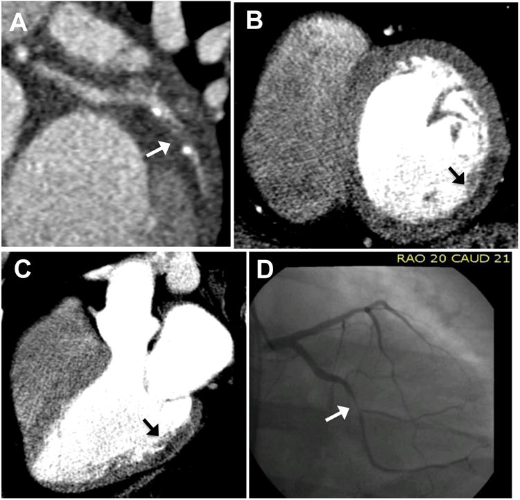
Coronary CT angiography reveals obstructive CAD (arrow) with severe stenosis of the left circumflex artery (A). Resting CT perfusion 8-mm thick MPR short-axis reconstruction demonstrates a subendocardial to mid-myocardial mid-basal inferolateral rest perfusion defect (arrow) (B) which is confirmed in the 3-chamber view (C). This 39-year-old man subsequently underwent SPECT-MPI which revealed an inferolateral fixed stress and rest defect with peri-infarct ischemia (not shown). Invasive coronary angiography (D) revealed a 95% left circumflex stenosis.