
Figure 3. Incremental value of resting CT Perfusion.
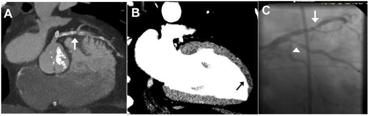
CTA images (A) yielded indeterminate coronary stenosis severity secondary to heavy calcification and slab artifact (arrow). Resting CT perfusion reconstruction via 2-chamber view 8-mm thick MPR (B) demonstrate an apical anterior rest perfusion defect (arrow). In this 64-year-old man, the standard care evaluation included a stress test which revealed inferior ischemia and transient ischemic dilatation, and subsequent invasive coronary angiography (C) revealed a 70% LAD stenosis (arrowhead) and a 90% ramus intermedius stenosis (arrow).