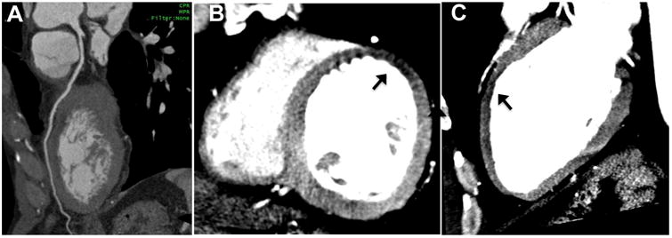
Figure 4. Discordance between coronary anatomy and resting CT perfusion.

Curved multiplanar reconstruction CTA image reveals no CAD along LAD (A), but resting CT perfusion analysis (B and C) using 8-mm thick MPR demonstrates a mid-anterior rest perfusion defect (arrows) in short-axis and 2-chamber views. In this 52-year-old man presenting with chest pain, serial troponin was mildly elevated leading to urgent invasive coronary angiography (not shown) which revealed mild narrowing of the LAD that resolved with intra-coronary nitroglycerin, consistent with coronary vasospasm.
