Fig. 2.

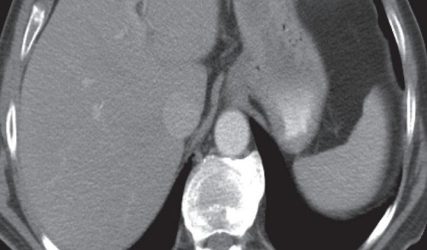
Contrast enhanced computed tomography axial image showing a well-circumscribed submucosal lesion along the lesser curvature of the stomach without necrosis.

Contrast enhanced computed tomography axial image showing a well-circumscribed submucosal lesion along the lesser curvature of the stomach without necrosis.