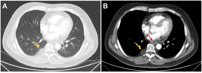
Figure 4.

Variations in CT in the ESCC 1 month after finishing all the treatments.
Note: (A and B) The stabilization of the right pleural mass (yellow arrows) and decrease of the thickened esophageal wall (red arrow) after the treatment, respectively.
Abbreviations: CT, computed tomography; ESCC, esophageal squamous cell carcinoma.
