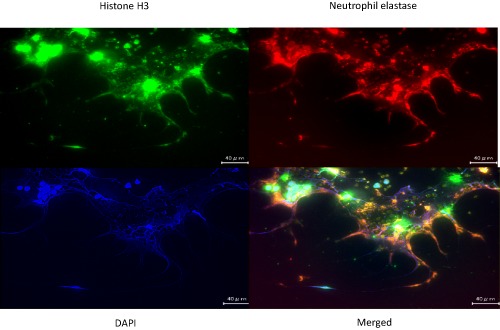
Figure 2.

Representative images showing fluorescent immunostaining of neutrophil extracellular traps (NETs). Neutrophils from a healthy volunteer were treated with phorbol myristate acetate for 4 h, then fixed with 4% formalin on the cover glass, and stained with three different colors: green, anti‐histone H3 monoclonal antibody; blue, DNA labeled with 4′,6‐diamidino‐2‐phenylindole (DAPI); red, anti‐elastase monoclonal antibody (objective: ×40). Scale bar = 40 μm. Original data.
