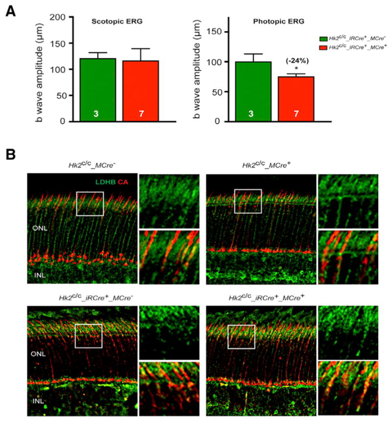
Figure 6. Retinal Function in HK2 Double Knockout Mice.

(A) ERG recordings in HK2 double knockout mice at 4 months of age showing no difference in the b-wave amplitudes of scotopic single-flash ERG responses at 0.01 cd*s/m2 between Hk2c/c_iRCre+ and Hk2c/c_iRCre+_MCre+ littermates. In contrast b-wave amplitudes of photopic responses show reduced cone function only in iRCre+_MCre+ retinas. Errors bars ± SD; numbers in bars, number of mice analyzed.
(B) Representative IHC images on retinal sections for LDHB expression (green signal) at 1 (top) or 2 (bottom) months of age. Cones were detected with an anti-cone arrestin antibody (CA, red signal). Higher magnification of boxed areas is shown to the right of each panel. INL, inner nuclear layer; ONL, outer nuclear layer.
