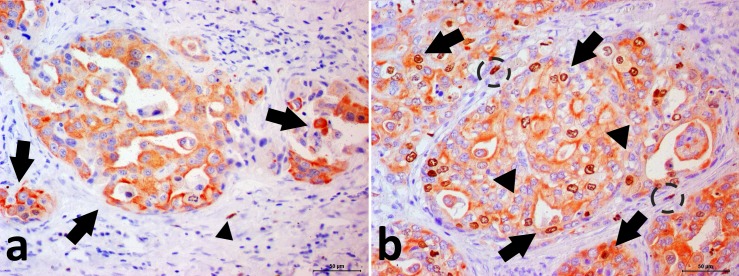
Fig 3. Photomicrograph showing the canine prostate cancer.
Double immunohistochemical staining for p63 and CK8/18. (A) PC with p63-/CK8/18+ phenotype. It is possible to note cytoplasmic CK8/18 expression (red staining—arrow) in neoplastic cells and nuclear negative p63 staining in neoplastic cells. There is a p63+/CK8/18- basal cell (arrowhead) (internal control). Negative signal can be observed as the blue color. (B) PC with p63+/CK8/18+ phenotype. Note the neoplastic doubled stained cells (arrows). It is also observed p63-/CK8/18+ cells (arrowhead) and p63+/CK8/18- basal cells (doted circle).

