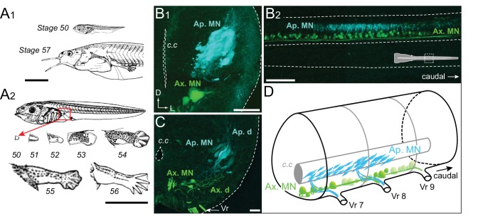
Figure 1. Developmental stages of X. laevis and anatomical organization of the spinal motor columns.
(A) Relative size differences between stage 50 and 57 larvae (A1) and associated morphological changes of the hindlimb bud during metamorphosis onset (A2), from Nieuwkoop and Faber (1956). (B, C) Segmental (B1, C) and rostrocaudal (B2) organization of the spinal motor column region containing limb MNs at stages 50–52 (B1, B2) and 55 (C). Inset in B2 shows the location of labeled MNs in the mid-region of the spinal cord. (D) Schematic representation of the segmental organization of the appendicular and axial motor columns in the larval spinal cord. Ap./Ax. MNs, appendicular/axial motoneurons; Ap./Ax. d., Ap./Ax. dendrites; Vr, ventral root; c.c, central canal; D, dorsal; L, lateral. Scale bars: A1 and A2 = 5 mm; B1 and C = 20 µm; B2 = 100 µm.

