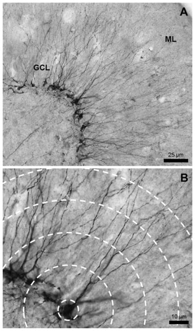
Figure 2.
Representative image of DCX+ immature neurons. (A) The DCX+ cell bodies line the inside of the granule cell layer (GCL) and the dendrites extend outward toward the molecular layer (ML). Image taken with a 20× lens. (B) Dendrites were traced at a high magnification which allows clear visualization of the processes and bifurcations. Image taken with a 40× lens.

