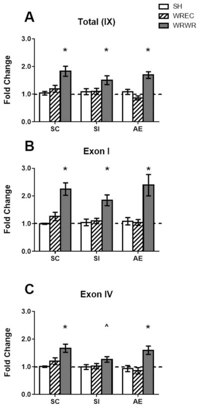
Figure 7.

Bdnf gene expression in the PD72 hippocampus. (A) WRWR showed more total Bdnf gene expression compared with SH and WREC animals. (B) Exon I- and (C) exon IV-specific transcripts were significantly elevated in all WRWR animals. A trend toward an increase was found for exon IV-specific gene expression in WRWR pups relative to SH (p <0.1, indicated aŝ on graph). * p <0.05. Data is expressed as a fold change from the SH-suckle control (SC) group (shown as 1 on graphs).
