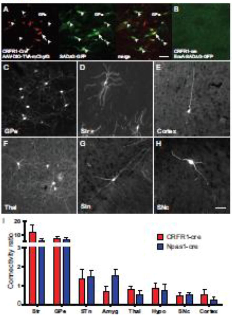
Figure 4. Rabies viral tracing of CRFR1+ and Npas1+ GPe neurons.
(A) Injection of AAV helper viruses encoding Cre-dependent TVA-mCherry (red) and G allow for infection by EnvA pseudotyped, G-deleted rabies virus that expresses GFP (SADΔG-GFP, green). The merged image shows a starter cell that expresses both the helper and the rabies viruses (yellow, arrow) surrounded by neurons expressing GFP only (green, arrowheads) which have been transynaptically infected via a synapse with a starter cell. Scalebar = 50µm. (B) Injection of pseudotyped rabies virus in animals without helper virus injection results in no infection, and no GFP expression. (C–H) Patterns of monosynaptic retrograde infection were broadly similar in CRFR1-cre and Npas1-cre experiments. The most presynaptic neurons were located in the GPe (C) and the Striatum (Str; D). We also observed neurons in the Cortex (E), Thalamus (Thal; F), Sub-thalamic nucleus (STN; G) and Substantia Nigra pars compacta (SNc; H). Quantification of tracing data reveals that projection patterns to CRFR1+ GPe neurons (red) and Npas1+ GPe neurons (blue) are broadly similar across many brain regions. n=3 per genotype.

