
Figure 1.
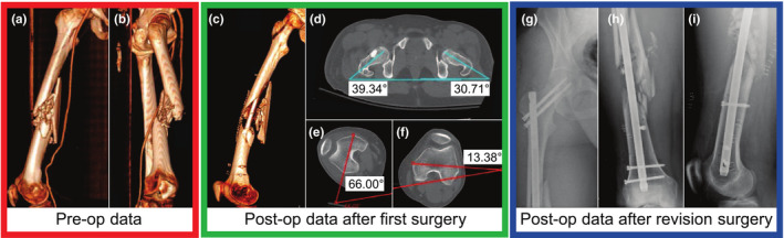
Difficulties arise in addressing rotational alignment in long bone fractures—the 3D preoperative CT scan of the right femur of a patient with a ballistic fracture of the femoral shaft is shown in (a, b). As seen in these images, due to the significant comminution, there are few anatomical cues as to the correct rotational alignment of the bone. (c) shows the postoperative CT of the same femur after reduction and placement of a cephallomedullary nail. The varus/valgus alignment appears to be restored (see Fig. 2); however, significant rotational malalignment is present with excessive external rotation of the distal aspect of the femur. Axial cuts from the postoperative CT scan are shown in (d–f). As shown in (d), the hips are in relatively similar position (right hip ∼10° externally rotated vs. the left). However, in (e), the operative right knee is over 40° more externally rotated than the healthy contralateral side in (f). Figures (g–i) show the anteroposterior (AP) view of the right hip, AP view of the right femur, and the lateral postoperative radiographs after revision cephalomedullary nailing with correction of the rotational deformity. The revision surgery includes removal and correct replacement of the intramedullary nail. [Color figure can be viewed at wileyonlinelibrary.com]