
Figure 2.
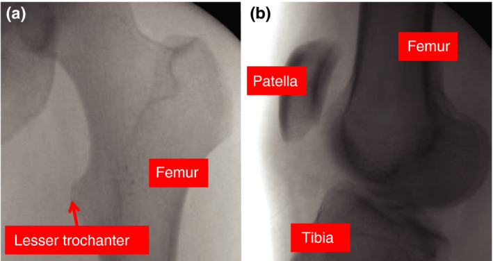
Contralateral images for guidance in rotational alignment—(a) and (b) are intraoperative fluoroscopic images from the revision surgery; AP view of the contralateral hip and lateral view of the contralateral knee. These images were utilized to guide rotational alignment of the fractured femur. By visualizing landmarks on these radiographs and understanding the change in angulation of the C‐arm, the surgeon can estimate the rotational alignment of the healthy femur and attempt to recreate this alignment on the operative side. [Color figure can be viewed at wileyonlinelibrary.com]