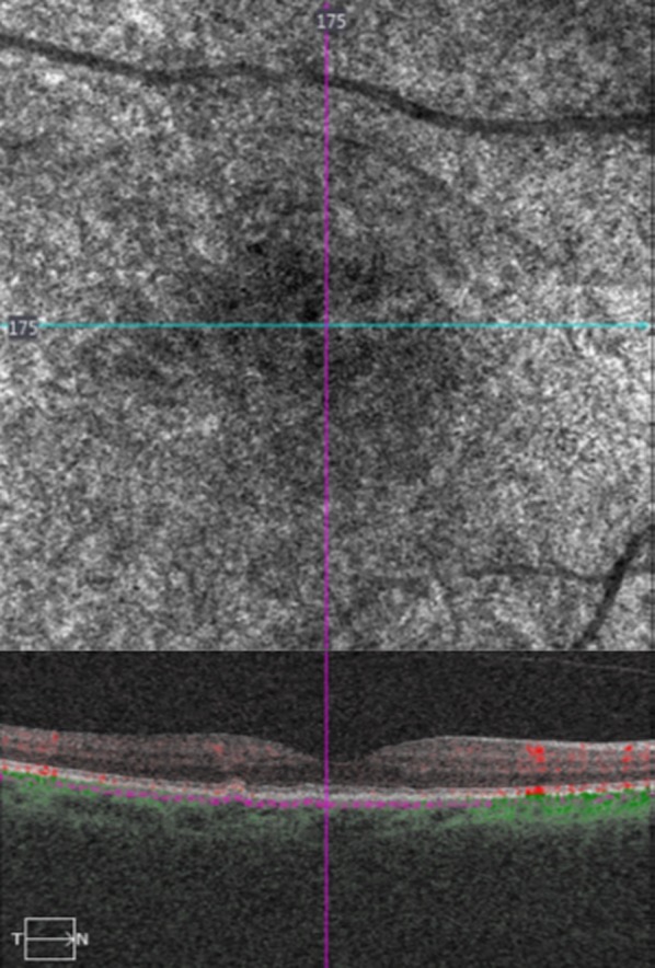
Fig. 3.

(Top) Optical coherence tomography angiography (OCTA) and (bottom) spectral domain optical coherence tomography (OCT) in a female patient with small drusen, showing almost normal choriocapillaris’ density, which was interrupted by small, black holes, correspondent to the small drusen area
