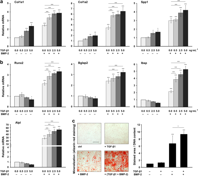
Fig. 4.
TGF-β1 exhibits a stimulatory and dose-dependent effect on BMP-2-induced osteoblast differentiation. a, b Effect of recombinant TGF-β1 and BMP-2 on the expression of osteoblast-specific genes in ST2 cells. Cells were treated with increasing concentrations (0.0, 0.5, 2.5, and 5.0 ng·mL-1) of TGF-β1 in the absence (−) or presence (+) of 100 ng·mL-1 BMP-2 for 24 h before total RNA was extracted. Expression of Col1a1, Col1a2, and Spp1 mRNAs (a), as well as Runx2, Bglap2, Ibsp, and Alpl mRNAs (b) was analyzed by qRT-PCR. Values normalized to Gapdh are expressed relative to the values of untreated cells. Mean± standard deviations were obtained from three independent experiments, and significant differences to either untreated cells or cells treated with BMP-2 alone (***P < 0.001, **P < 0.01, *P < 0.05) are shown. c Effect of the recombinant TGF-β1 and BMP-2 on the mineral deposition capacity of ST2 cells. Cells were treated with TGF-β1 (5 ng·mL-1), BMP-2 (100 ng·mL-1), or both growth factors for 4 d, and were subsequently grown under osteogenic culture conditions for 10 additional days before extracellular matrix mineralization was assessed by alizarin red staining. Representative images are shown. Scale bar=500 µm. Mineral deposition capacity was quantified by measuring the stained area using the Fiji distribution of ImageJ. Values normalized to the DNA content are expressed relative to the values of untreated control cells (ctrl). Data and statistical significance are expressed as in a, b

