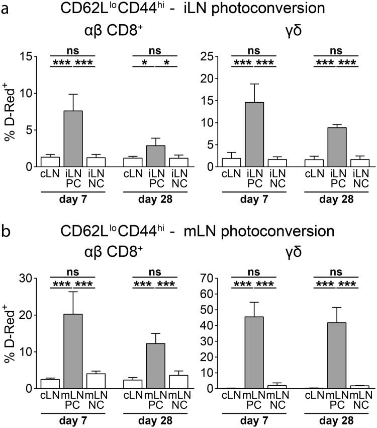
Figure 4.
Peripheral and mesenteric LNs harbor resident γδ T cells. (a) Frequency of D-Red+ cells among CD62LloCD44hi among αβ CD8+ T cells (left) and γδ T cells (right) in the indicated LNs 7 and 28 days after photoconversion of iLN (n = 4–6 mice per time point in 5 independent experiments, mean ± SD, one-way ANOVA with Tukey’s multiple comparisons test, *P < 0.05; ***P < 0.001; ns: not significant). (b) Frequency of D-Red+ cells among CD62LloCD44hi for αβ CD8+ T cells (left) and γδ T cells (right) in the indicated LNs 7 and 28 days after photoconversion of mLN (n = 4–5 mice per time point in 5 independent experiments, mean ± SD, one-way ANOVA with Tukey’s multiple comparisons test, ***P < 0.001; ns: not significant).

