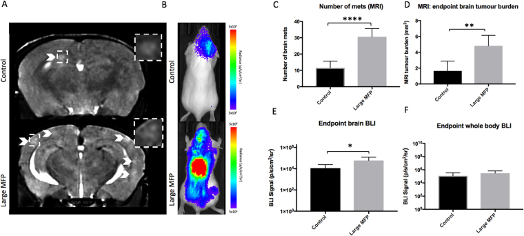
Figure 4.
(A) At day 14, brain metastases appeared as regions of hyperintensity by MRI, and (B) regions of BLI signal in the brain and body. (C) Mice with a large MFP tumor had significantly more brain metastases than mice without a primary tumor. (D) Mice with a large MFP tumour had significantly more total brain tumor burden than mice without a primary tumor. (E,F) Similarly, mice with a large MFP tumour had significantly more BLI signal in both the brain and the body compared to control mice. Data is presented as mean +/− SD. *Indicates p < 0.05; **indicates p < 0.01.

