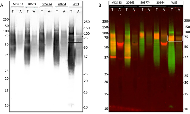
Figure 3.
A-LPS modification of PPAD in sorting type I and II isolates of P. gingivalis. Cells of P. gingivalis sorting type I and II isolates were cultured in BHI and, subsequently, extracted with Triton X-114. Upon phase separation at 37 °C, proteins in the detergent-rich (T) and aqueous (A) phases were analyzed by LDS-PAGE and Western blotting. In panel A immunodetection was performed with an A-LPS-specific monoclonal antibody. Panel B shows a dual-labeling image upon immunodetection with the A-LPS-specific monoclonal antibody (green signal) and a PPAD-specific rabbit antibody (red signal). Bands representing the A-LPS-modified PPAD species of ~75–85-kDa PPAD are boxed in both panels. Names of sorting type I isolates are underlined. Molecular weights of marker proteins are indicated. The full-length blot is presented in Figure S1. Please note that the order of ‘A’- and ‘T’-labeled lanes is inversed compared to Fig. 2 as samples were loaded in a different order.

