
Figure 5.
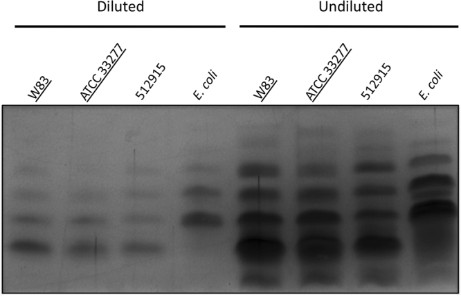
LPS fingerprints of PPAD sorting type I and II isolates. Total LPS was extracted from P. gingivalis sorting type I and II isolates, and an E. coli control. Subsequently, the extracted LPS was separated by SDS-PAGE and visualized by silver staining. To detect possible differences in the fingerprints, extracted LPS was loaded in a ten-fold dilution and undiluted. Names of sorting type I isolates are underlined. The full-length gel picture is presented in Figure S1.