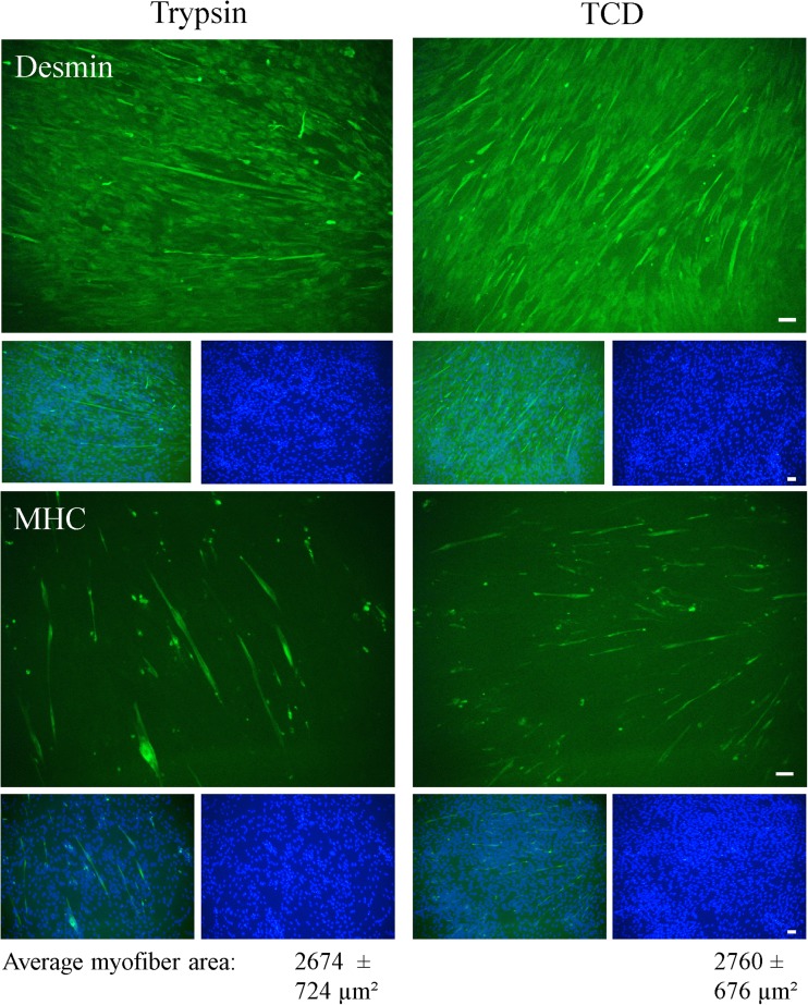
Figure 2.
Differentiation potential of SC/MPC liberated with trypsin alone or with TCD. Isolated cell populations were seeded on Matrigel-coated plates at days 3–6 after isolation, and first cultured in growth medium. Subconfluent cells (about 80%) were transferred to differentiation medium after 4 to 5 d to induce myogenic differentiation. After 5 d in differentiation medium, cells were fixed and immunostained for Desmin and MHC to indicate their differentiation potential; cell nuclei were stained with DAPI. Quantitative analysis of differentiation was performed by encircling MHC+ myotubes (≥ 2 nuclei) to determine myotube area and results are illustrated in the figure as mean ± SD. SC/MPC dissociated with trypsin and TCD were both able to initiate myogenic differentiation (MHC+ cells) and started to form multinucleated myotubes. The images are representative of three individual experiments (n = 3 piglets) and 5–6 ramdom sections were evaluated per experiment. Scale bar represents 50 μm.

