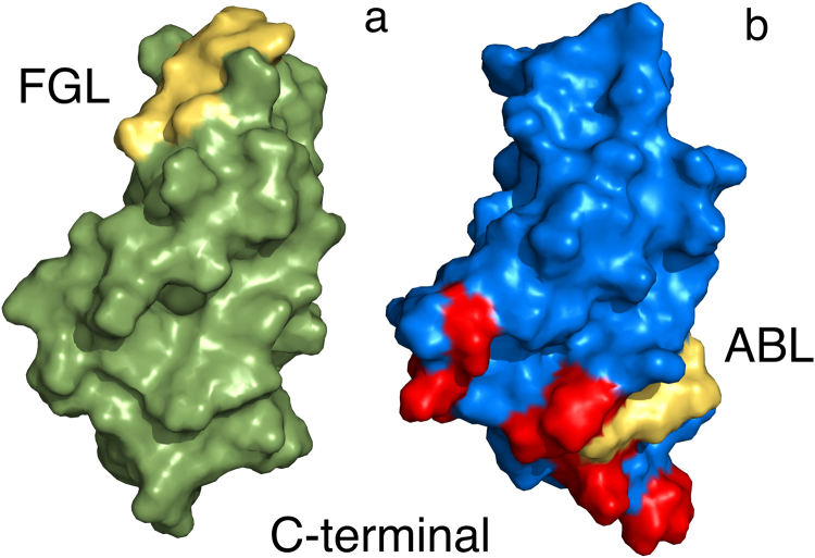
Figure 1.
The Walker A motif is located differently in NCAM1 and NCAM2. The figure shows the locations of the Walker A motif in the FnIII2 domains of NCAM1 ((a) PDB code: 1lwr) and NCAM2 ((b) PDB code: 2kbg). The domains are presented as surface models, and are oriented in a manner that enables the visualization of the Walker A motifs, which are located differently in the two proteins. Moreover, the domains are oriented with the membrane-proximal end facing down. FGL and ABL indicate the Walker A motif-containing loop regions of NCAM1 and NCAM2, respectively. The Walker A motifs and residues perturbed when titrating NCAM2 FnIII2 with AMP-PCP are colored yellow and red, respectively.

