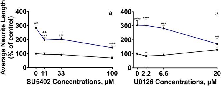
Figure 5.
NCAM2 FnIII1-2 activates signaling through FGFR and MEK. CGNs were treated for 24 h with different concentrations of the FGFR kinase inhibitor SU5402 (Sigma Aldrich; a) or the MEK inhibitor U0126 (MERCK; b) in the absence (black curves) or presence (blue curves) of 4 μM NCAM2 FnIII1-2. The inhibitors were added to the wells 10 min after the cells had been plated. In each experiment, 4 concentrations were used per inhibitor: 0, 11.1, 33.3 and 100 μM (for SU5402), and 0, 2.2, 6.6 and 20 μM (for U0126). Average neurite lengths were estimated by a stereological approach from recorded micrographs. Data are shown normalized to control (0 μM inhibitor in the absence of FnIII1-2) as mean and SEM (n = 3). The individual concentration-response data were analyzed by one-way ANOVA for repeated measures followed by Tukey’s multiple comparison test. (a) F (12, 35) = 36.55, p < 0.0001. (b) F (12, 35) = 28.14, p < 0.0001. *,** and ***p < 0.05, 0.01, and 0.001, respectively, when compared to negative control (no FnIII1-2, 0 μM inhibitor). +,++ and +++p < 0.05, 0.01, and 0.001, respectively, when compared to positive control (FnIII1-2, 0 μM inhibitor). The effect of the total average neurite length per cell for domains cultured with inhibitor can be found in Supplementary Table S2.

