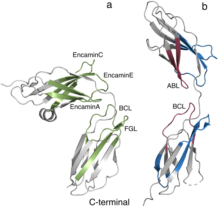
Figure 6.
Structural comparison of the NCAM1 and NCAM2 FnIII domains. The FnIII domains (grey) from NCAM1 ((a) PDB code: 2vkw) and NCAM2 ((b) PDB code: 2jll) were aligned to compare secondary structure differences. The domains are oriented with the membrane-proximal end facing down. Pairwise, the domains have same overall topology. However, NCAM1 FnIII1 contains an α helix between the D and E strands not observed in NCAM2 FnIII1. Biologically active peptides derived from NCAM1 FnIII1-2 (EncaminA, -C, -E, BCL and FGL) are colored in green. All NCAM1 FnIII1-2 peptides are in the literature reported to interact with FGFR53. Peptides derived from NCAM2 FnIII1-2, and used in this study are colored red (biologically active, ABL and BCL) and blue (not biologically active). When comparing the regions of NCAM1, from which the respective peptides are derived, with the corresponding regions in NCAM2, they all have highly identical secondary structures.

